

Vertebral segmentation & alignment
Spinal alignment, measured automatically.
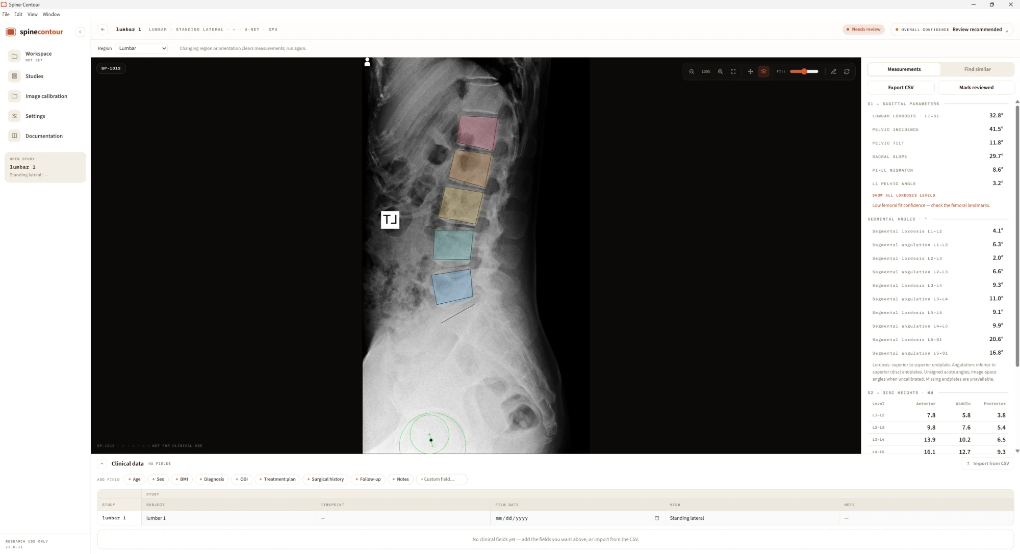
SpineContour is an open-source research application that measures cervical, lumbar and standing full-spine radiographs, detecting which it has been given. It reports the alignment parameters used in spine research: C2–C7 Cobb angle and cervical SVA, lumbar lordosis, pelvic incidence, pelvic tilt, sacral slope, PI–LL mismatch and L1 pelvic angle, and the global C7–S1 SVA on standing films. It runs in seconds, entirely on your workstation.
01 · What it does
Segmentation and landmarks
Contours for L1–S1 with a per-study confidence score, and 23-point landmarks on cervical films. Review and correct either directly on the image.
Alignment parameters
C2–C7 Cobb and cervical SVA, lumbar lordosis, pelvic incidence, pelvic tilt, sacral slope, PI–LL mismatch and L1 pelvic angle, plus segmental lordosis and disc angulation at every level. T1 slope and T4 pelvic angle once you place T1 or T4 by hand. Exportable as CSV or a PDF report.
Local by design
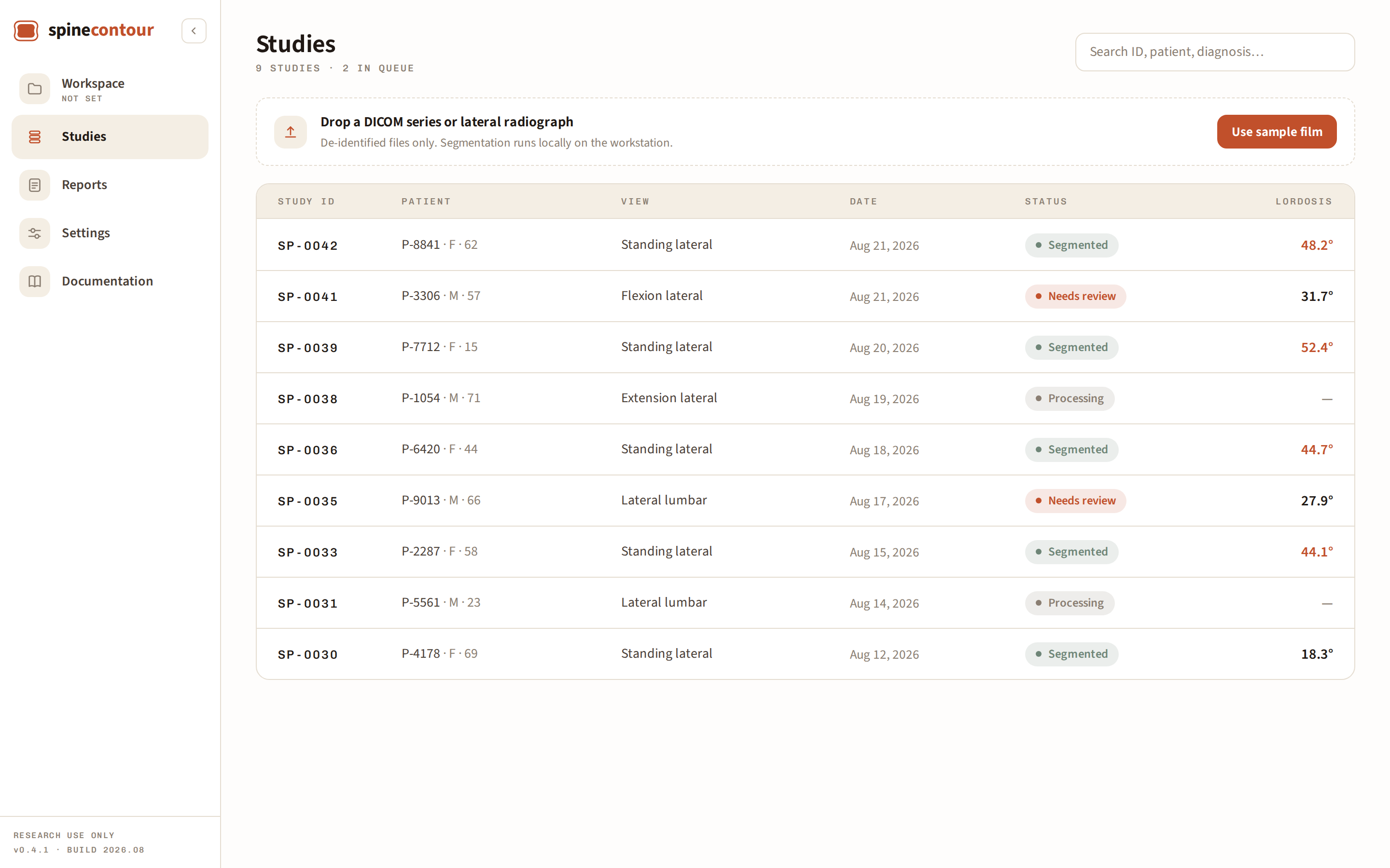
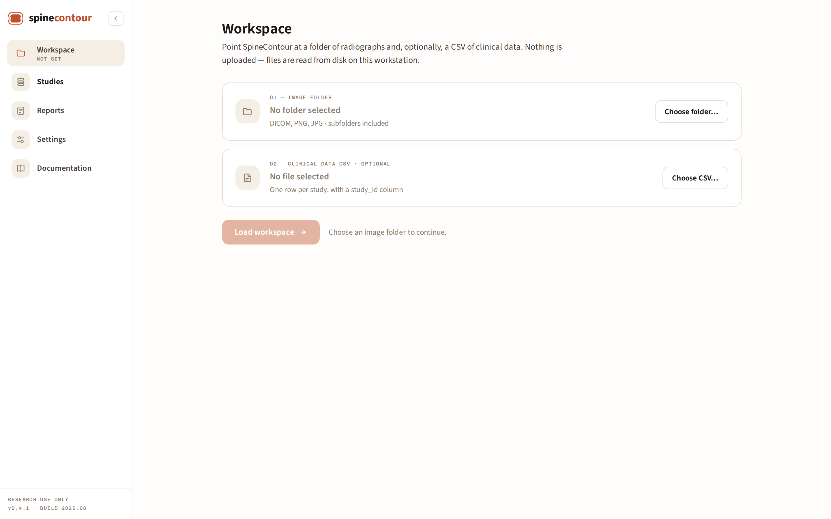
Nothing is uploaded. Files are read from disk on this workstation, and de-identified research images never leave your machine.
Your data never leaves your workstation
SpineContour runs entirely on the machine you install it on. Radiographs and clinical data are read from disk and stay there. There is no mechanism in the application to send them anywhere: no upload, no cloud processing, no telemetry, no account.
- No upload path for images, measurements, or reports
- No account, login, or license server
- No analytics, telemetry, or crash reporting
- Clinical data is imported from a CSV file you choose
- Exports are written to a location you pick on disk
- Works with networking disabled
02 · Inside the app

03 · Measurement accuracy
Externally validated against expert measurement.
Four networks were trained on 10,387 images from four public datasets: three HRNet models for the vertebrae (C2–C7 and L1–S1 endplates) and a binary U-Net for the femoral heads. Testing used 100 lateral full-spine radiographs from the Lateral Full-spine X-ray Image Dataset, a separate collection excluded entirely from model development. Uncorrected automated measurements were compared with a neurosurgery spine fellow's reference measurements by mean absolute error, root mean square error, prediction R², and absolute-agreement ICC(A,1).
| Parameter | ICC(A,1) | Agreement |
|---|---|---|
| Global SVA | 0.998 | Excellent |
| Pelvic tilt | 0.983 | Excellent |
| C2–C7 SVA | 0.960 | Excellent |
| Sacral slope | 0.933 | Excellent |
| Pelvic incidence | 0.916 | Excellent |
| C2–C7 Cobb angle | 0.835 | Good |
| L1–L5 lordosis | 0.834 | Good |
Disc heights: MAE 0.68 mm (C2–C3, middle) to 2.83 mm (L4–L5, anterior). Segmental angulation: MAE 2.70° (C4–C5) to 4.36° (L2–L3). Images with a missing landmark, such as an obscured C7, were not evaluated on the parameters that depend on it. Full MAE, RMSE and R² results are in the forthcoming methods paper.
Not yet validated: L1 pelvic angle, T4 pelvic angle and T1 slope are reported by the app but have not been validated, because no reference dataset with these measurements is available to us yet. Treat them as exploratory.
04 · Research use
Not for diagnostic or clinical use.
SpineContour is an investigational research tool. Its measurements, segmentations, and derived metrics have not been cleared or approved by any regulatory body. All output requires independent verification by a qualified reviewer.
The model weights, training and evaluation code, and annotation tool are released openly. We invite other centers to test the platform, use it in their own research, and contribute annotations so that accuracy can be established across sites.
We're committed to keeping SpineContour open source and free to use. Please contact spine-contour@gmail.com with questions or if you'd like to collaborate.
spine-contour: An Open Source Platform for Automated Spine Segmentation and Spinopelvic Parameter Measurement (methods paper · forthcoming)
Datasets used in model development · 10,387 images
BUU-LSPINE
Lateral lumbar radiographs with published vertebral endplate coordinates, from Burapha University Hospital.
VinDr-SpineXR
Full-spine radiographs from two hospitals in Hanoi. Together with Merlin, contributes manually annotated images adding vertebroplasty cement and posterior instrumentation examples.
Merlin
Abdominal CT volumes from a United States academic center, released with the Merlin vision–language foundation model.
05 · Get SpineContour
Download SpineContour.
Version 1.6.1. Builds are published on GitHub, each with its checksum, so the link here is always the current release.
Neither build is code-signed yet, so your machine will warn you the first time.
- Windows shows a blue "Windows protected your PC" screen. Choose More info, then Run anyway.
- macOS refuses to open an unidentified developer. Control-click the app and choose Open, or allow it under System Settings, Privacy & Security.
The macOS build is for Apple Silicon only. Intel Macs are not supported.
SHA-256 · v1.6.1
Spine-Contour-Windows.exe
793cd51008597ef123c64518f27c2c4fb4d6df590e8179953a5ab5014a711447
Spine-Contour-macOS-arm64.dmg
0e15f51d029adc991be4495b71ed15d1a80001dd85ac68181124ae91f5785a9b
Check yours with
Get-FileHash .\Spine-Contour-Windows.exe in PowerShell, or
shasum -a 256 Spine-Contour-macOS-arm64.dmg in Terminal.